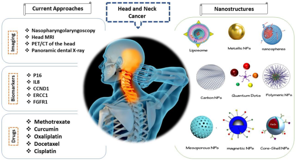
| Diagnosis of head and neck cancer typically begins with a thorough medical history and physical examination, including a visual inspection of the mouth, throat, and neck. Further investigations may include imaging techniques like CT scans, MRI scans, and PET scans to identify the tumor's location, size, and extent of spread. A biopsy, involving the removal of a tissue sample for microscopic examination, is crucial for confirming the diagnosis and determining the type and grade of cancer. Additional tests such as blood tests and endoscopy may also be performed to assess overall health and the stage of the cancer. 
Diagnosing head and neck cancer involves a multi-step process that typically includes:
1. Medical History and Physical Examination: The doctor will begin by taking a detailed medical history, including any risk factors (e.g., tobacco use, alcohol consumption, HPV infection, family history), symptoms (e.g., persistent sore throat, lump in the neck, difficulty swallowing, hoarseness), and duration of symptoms. A thorough physical examination of the head and neck region will follow, looking for any visible abnormalities like lumps, ulcers, or changes in the tissues.
2. Imaging Tests: Several imaging techniques help visualize the tumor and its extent:
- Panoramic X-ray: A standard X-ray of the jaw and teeth, often used to assess bone involvement.
- Computed Tomography (CT) scan: Provides detailed cross-sectional images of the head and neck, allowing for precise localization of the tumor and assessment of its size, location, and spread to nearby structures.
- Magnetic Resonance Imaging (MRI) scan: Offers superior soft tissue contrast compared to CT, particularly helpful in evaluating the involvement of nerves, muscles, and blood vessels.
- Positron Emission Tomography (PET) scan: Used to detect cancer cells throughout the body, helping to determine if the cancer has metastasized (spread). Often combined with a CT scan (PET/CT).
3. Biopsy: This is the most crucial step for definitive diagnosis. A small tissue sample is taken from the suspicious area and examined under a microscope by a pathologist. There are various biopsy techniques, including: - Fine-needle aspiration biopsy (FNAB): A thin needle is used to collect cells.
- Incisional biopsy: A small piece of the tumor is removed.
- Excisional biopsy: The entire tumor is removed.
The biopsy determines the type of cancer cells, their grade (how aggressive they are), and other important characteristics.
4. Endoscopy: A thin, flexible tube with a camera is used to visualize the upper respiratory and digestive tracts. This can help identify tumors that are not easily visible during a physical examination. Examples include: - Nasopharyngoscopy: Examination of the nasopharynx (back of the nose and throat).
- Laryngoscopy: Examination of the larynx (voice box).
- Esophagoscopy: Examination of the esophagus (food pipe).
5. Staging: Once the diagnosis is confirmed, the cancer is staged. Staging involves determining the size and extent of the tumor, whether it has spread to nearby lymph nodes or distant organs, and its overall impact on the patient's health. The TNM system (Tumor, Node, Metastasis) is used to classify head and neck cancers based on these factors. Staging is essential for treatment planning and prognosis.
Important Note: This information is for general knowledge and should not be considered medical advice. If you suspect you may have head and neck cancer, it's crucial to consult a medical professional for proper diagnosis and treatment. Early diagnosis significantly improves treatment outcomes.
Tags: CT Scans Endoscopy HPV Infection MRI Scans Nanostructures Neck Cancer PET Scans  
|  1,221
1,221  0
0  0
0  3350
3350 